
The Link Between COVID-19, mRNA Vaccines and Vitamin A

COVID-19 vaccines have been associated with an unusually high number of adverse events (VAEs) and deaths compared to other vaccines and medicinal products in general. At the time of writing, 12,131 reports of myocarditis or pericarditis have been submitted to the US VAE Reporting System (VAERS), in addition to 9,094 reports of myocardial infarction and 18,461 reports of fatalities following vaccination against COVID-19.
mRNA vaccines introduce mRNA of a modified SARS-CoV-2 spike glycoprotein (S). S has recently been shown to bind to receptor for retinol uptake STRA6 (STRA6) with a greater affinity than to angiotensin-converting enzyme 2 (ACE2), and it is believed to inactivate STRA6 (Mahmoud, Tamer et al. 2021). These findings give rise to speculation that some or many COVID-19 symptoms and VAEs could be the result of decreased retinyl ester biosynthesis and storage, and increased diffusion of unbound retinol into tissues.
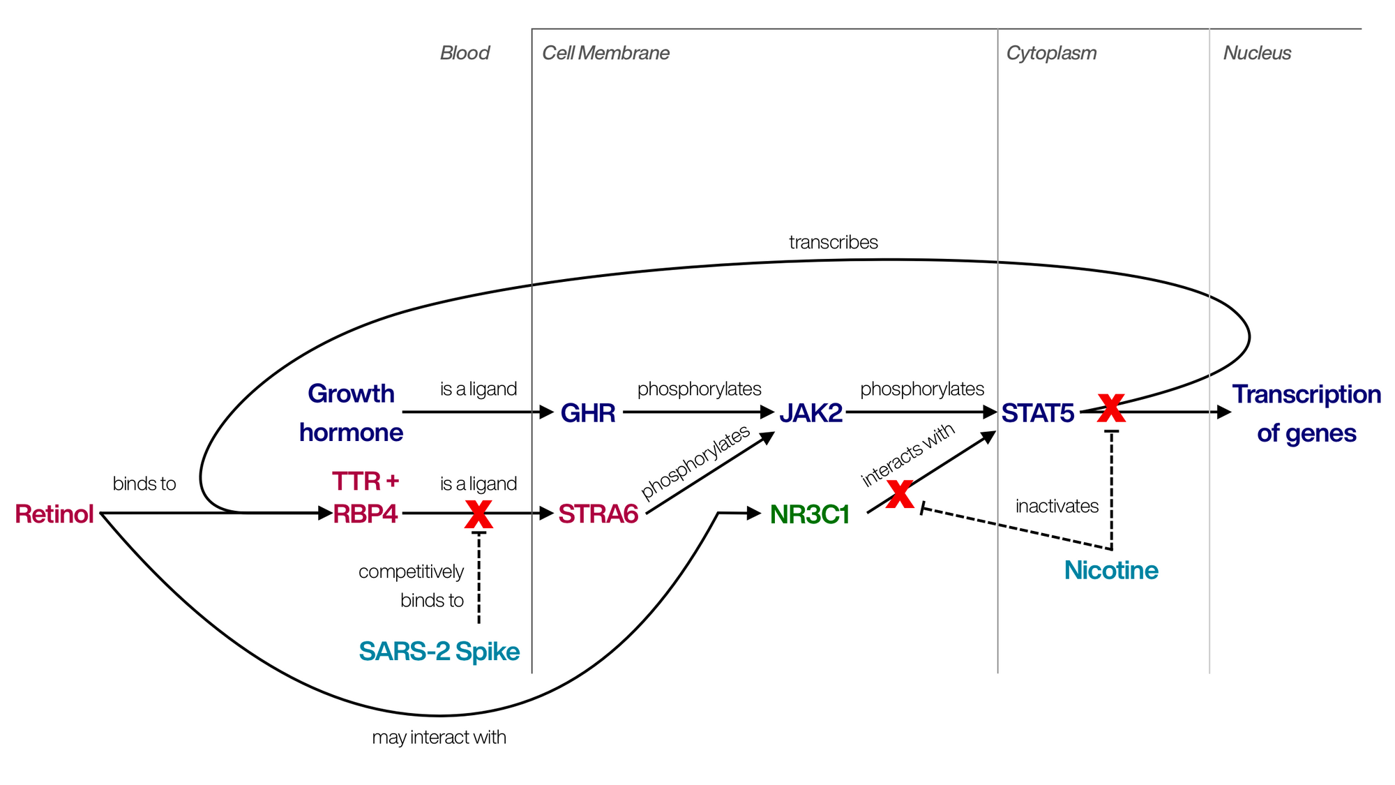
Inactivation of STRA6 prevents cells from accepting retinol bound to retinol-binding protein 4 (RBP4) and therefore also prevents esterification of retinol by lecithin retinol acyltransferase (LRAT; Marwarha, Berry et al. 2014). Furthermore, since signaling by STRA6 leads to phosphorylation of signal transducer and activator of transcription 5A (STAT5A), inactivation of STRA6 is expected to decrease transcriptional activity of STAT5. Because retinol induces its own metabolism, inactivation of STRA6 likely also results in a decrease of serum RBP4, even though the binding of retinol to RBP4 is crucial to prevent retinoid toxicity. Furthermore, osteoporosis is the condition most commonly associated with vitamin A overdose (Hough, Avioli et al. 1988), and mice surviving SARS-CoV-2 infection were recently found to exhibit a 24% decrease in bone mass and 64% increase in osteoclasts (Awosanya, Dalloul et al. 2022). Analyzing the expression of vitamin A-related genes after vaccination will help clarify the role of vitamin A in both COVID-19 and VAEs.
For this investigation I analyzed the expression of genes in plasma samples taken before, and on different days after administration of both COMIRNATY® (Pfizer-BioNTech COVID-19 Vaccine) doses, using the data published by (Arunachalam, Scott et al. 2021). Expression of genes on days 1, 2, 7, 21, 22, 28 and 42 post-vaccination was compared to the expression of genes before vaccination (i.e. day 0). The second vaccine dose was administered on day 22 (D22). The data is from 6 participants, although day 1-data was missing for 3 of the 6 participants. Therefore, the results for D1 are less significant than the remaining results. Additionally, based on the expression of Y-linked genes, the results for D1 appear to be from male participants only.
Methods
Gene expression calculations and generation of figures was performed using an app developed for this purpose. Results are expressed as percent change in expression of the gene in a test group compared to the expression of the same gene in the control group. For example, a value of 0% is equivalent to a fold-change of 1 or a log2(fold-change) of 0, and a value of -50% is equivalent to a fold-change of 0.5 or a log2(fold-change) of -1. Zero or negative values were excluded and assumed to be non-quantifiable. Error bars in the generated charts denote the standard deviation.
Significance of the results was determined for each group versus control, and additionally for each group versus all other test groups, using Welch’s t test, which was reported to be reliable for this purpose (Ullah, Paul et al. 2019). (*), (**) and (***) denote a significant change versus control with p < 0.05, p < 0.01 and p < 0.001, respectively. Correlations were established by calculating the Pearson correlation coefficient (r).
Non-quantifiable genes
mRNA of RBP4, which is secreted by the liver and adipocytes (Höpfinger, Berghoff et al. 2021), was only quantifiable in insignificant amounts, as was mRNA of STRA6 and transthyretin (TTR).
JAK2 is significantly upregulated after dose 2
Expression of tyrosine-protein kinase JAK2 (JAK2), which is attached to STRA6, was up nonsignificantly (+62%) after the first dose, and further increased after the second dose (+100%; p < 0.01) and on the following day (+46%; not significant). This seems to indicate that the interaction of S with STRA6 does not cause phosphorylation of JAK2, and that cells may increase expression of JAK2 to compensate for its lack of activity. Expression of glucocorticoid receptor (NR3C1), which was shown to interact with STAT5 (Engblom, Kornfeld et al. 2007), is also increased significantly after the second dose (+22%; p < 0.05). N-myc-interactor (NMI), which potentiates the activity of STAT family transcription factors (Zhu, John et al. 1999), was extremely correlated with JAK2 (r = 0.88) and concomitantly upregulated (D22: +76%, p < 0.01; D28: +43%, p < 0.05).
Also of note is suppressor of cytokine signaling 3 (SOCS3), which binds to and thereby deactivates JAK2 and was down after the first dose (-52%; p < 0.01) but up after the second dose (+78%; p < 0.01) and the following day (+41%; p < 0.05; data not shown). SOCS2 was up after the first dose (+55%; p < 0.01) but normalized thereafter and was unaffected by the second dose.
Signal transducer and activator of transcription 1 (STAT1), STAT2, STAT3 and STAT5 are significantly upregulated after vaccination
STAT1 is activated by interferon gamma (IFNG) when it binds to interferon gamma receptor 1 (IFNGR1). One of STAT1’s target genes is guanylate-binding protein 5 (GBP5), which activates pattern receptors in the innate immune system.
Expression of IFNG is up after the first dose (+151%; p < 0.001), less affected after the second dose but unexpectedly spikes again on D28 (+107%; p < 0.05). The full significance of STAT1 activation remains to be determined.
STAT2 is activated during a sequence of events initiated by interferon beta (IFNB1), which was not quantifiable in plasma. After phosphorylation by non-receptor tyrosine-protein kinase TYK2, or by JAK1, STAT2 dimerizes with itself or STAT1 and forms a complex with interferon regulatory factor 9 (IRF9). This complex induces transcription of genes from the IFN-stimulated response element (ISRE), and one of the target genes is interleukin-6 (IL6; Nan, Wang et al. 2018). IL6 is reported to play a major role in the pathogenesis of cytokine storm and the progression of COVID-19 (Giannakodimos, Gkountana et al. 2021).
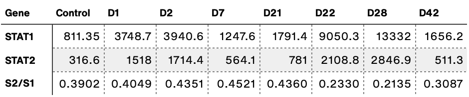
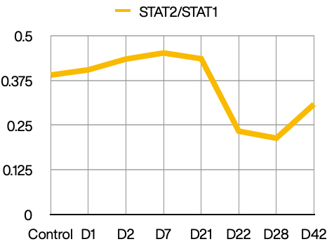
Very surprisingly, IL6 is extremely downregulated following the first dose (-61%; p < 0.05). Its expression normalizes thereafter and is upregulated after the second dose (+84%; p < 0.05). Expression of STAT2 was extremely correlated with STAT1 (r = 0.959), and levels of STAT2 mRNA were generally around 1.5x lower than levels of STAT1 mRNA. STAT2 was also highly correlated with IRF9 (r = 0.868), which was up after the second dose (+76%; p < 0.001).
Activation of STAT5 via retinol/STRA6 (Berry, Jin et al. 2011) or via growth hormone receptor (GHR; Liu, Nie et al. 2021) is believed to induce transcription of lipogenic genes, although it is unclear whether this can be achieved by STAT5 homodimers or whether peroxisome proliferator-activated receptor gamma (PPARG)/STAT5 heterodimers are required (Dentelli, Trombetta et al. 2009).
Expression of STAT5A was up after the first dose (+29%; p < 0.05) and after the second dose (+36%; p < 0.01), and did not decrease until D42 (+8%). STAT3 is activated in response to specific cellular damage that results in the liberation of certain substances including collagen, diglycerides and retinoic acid (RA). Expression of STAT3 was unaffected by the first dose but up significantly after the second dose (+44%; p < 0.001). Expression of STAT4 and STAT5B did not change significantly following vaccination (data not shown) while STAT6 only increased after the second dose (+19%; p < 0.05).
Vaccination differentially affects expression of protein kinases C (PRKC)
Proteins of the protein kinase C (PRKC) family transduce signals in a number of different pathways, and most of the PRKC members are inactivated by retinoic acid to variable degrees (Radominska-Pandya, Chen et al. 2000). PRKC alpha type (PRKCA) is capable of activating the MAPK/ERK pathway by phosphorylating RAF proto-oncogene serine/threonine-protein kinase (RAF1). PRKCB is activated by diacylglycerol (DAG) and in turn activates the canonical NF-κB pathway. PRKCE phosphorylates STAT3 at Tyr705, inducing dimerization and translocation to the nucleus (Shi, Papay et al. 2012). Therefore, inactivation or downregulation of PRKCE leads to a relative increase in Ser727-phosphorylated STAT3 (P-STAT3 Ser), which translocates to mitochondria instead of nucleus (Park, Lin et al. 2016).
Interestingly, PRKCA was persistently downregulated (-17 to -55%) after administration of the first vaccine dose, and the second dose had no apparent effect on expression of PRKCA. Expression of PRKCB and PRKCE was highly correlated (r = 0.807) and both were up following the second dose (PRKCB: +29%, p < 0.001; PRKCE: +33%, p < 0.05). PRKCD usually correlated with PRKCB and PRKCE, except on the day of the first dose (+41%; p < 0.05). Of the tested PRKCs, PRKCB was most abundant while PRKCE was least abundant.
Expression of prostaglandin G/H synthase 1 (PTGS1), which metabolizes all-trans-retinoic acid (atRA) to the pro-inflammatory (4S)-OH-RA, was up significantly after the first dose (+42%; p < 0.05), on D7 (+26%; p < 0.01), after the second dose (+47%; p < 0.001) and on D28 (+23%; p < 0.05). Alarmingly, expression of PTGS1 remained significantly elevated 42 days after the initial dose (+33%; p < 0.01). This finding is significant because PTGS1 also plays a role in blood clotting. PTGS1-null mice do not exhibit any major abnormalities except reduced inflammatory response after topical challenge with eicosatetraenoic acid (ETE) and decreased platelet activation, which may lead to increased bleeding and decreased clotting (Palma-Barqueros, Bohdan et al. 2021). Therefore, conversely, upregulation of PTGS1 could very likely produce blood clots similar to those reported after COVID-19 vaccination, and it could likely also produce the micro-clots observed in patients suffering from post-acute sequelae of COVID-19 (PASC; Pretorius, Vlok et al. 2021).
It should be noted that even though AstraZeneca has been associated with blood clots in media reports, blood clots reported to adverse events databases were caused by mRNA vaccines more often than by Vaxzevria. Furthermore platelet factor 4 (PF4), which has been linked to blood clots caused by Vaxzevria (Schultz, Sørvoll et al. 2021), was significantly upregulated following vaccination with Comirnaty, most prominently after the second dose (+106%; p < 0.001), and alarmingly also on D42 (+85%; p < 0.01). There was a modest correlation between PTGS1 and PF4 (r = 0.54).
Expression of retinol dehydrogenase (RDH) enzymes was only mildly affected by vaccination
Of the retinol dehydrogenase (RDH) family, only RDH11 and RDH14 differed significantly after vaccination. RDH8, RDH12 and RDH16 were not quantifiable. RDH11 was up after the first dose (+29%; p < 0.01), on D7 (+22%; p < 0.01) and after the second dose (+13%; p < 0.05), while RDH14 was up after the first dose (+16%; p < 0.05). RDH11, which reduces retinal to retinol under physiological conditions, was, together with RDH14, the most abundant RDH in plasma. None of CYP1A1, CYP2C8, CYP2C9, CYP2C18, CYP2C19, CYP7A1, CYP7B1, the CYP26s and BCO1 were quantifiable. Expression of beta,beta-carotene 9’,10'-oxygenase (BCO2) was not significantly changed although it increased after the initial dose (+48%). Expression of retinoic acid receptor RXR-alpha (RXRA) was up after the second dose (+25%; p < 0.05) while RXRB was up after the first dose (+21%; p < 0.05) and RXRG was not quantifiable. Fatty acid-binding protein 5 (FABP5), which transports retinoic acid and prefers 9-cis-RA over atRA, was consistently upregulated, most prominently after the second dose (+67%; p < 0.001) and on D28 (+54%; p < 0.05), while FABP4, which prefers atRA over 9-cis-RA was not quantifiable.
Of the hydroxysteroid 17-beta dehydrogenases (HSD17Bs), only HSD17B8, HSD17B10, HSD17B11 and HSD17B12 were quantifiable. Expression of HSD17B10, which catalyzes oxidation of (3S)-hydroxyacyl-CoAs to 3-oxoacyl-CoAs, was most upregulated after the second dose (+28%; p < 0.05), contrasting with HSD17B8, which was up on D21 (+22%; p < 0.05) but decreased after the second dose (+7%; not significant). Expression of very-long-chain 3-oxoacyl-CoA reductase (HSD17B12), which I have proposed to reduce 4-oxo-RA to the anti-inflammatory (4R)-OH-RA, was up on D7 (+17%; p < 0.05), after the second dose (+14%; p < 0.05) and on D28 (+13%; p < 0.01). The other HSD17Bs were not significantly affected.
Of the ALDH1As, which oxidize retinal to retinoic acid, only ALDH1A1 was quantifiable. ALDH1A1 was down nonsignificantly after the first dose (-15%) but up significantly following the second dose, reaching +83% (p < 0.05) and recovering by D42.
Evidence of hypoxia following vaccination
Hypoxia-inducible factor 1-alpha (HIF1A), which is induced by hypoxia, was downregulated nonsignificantly following dose 1 (-27%) but up after dose 2 (+72%; p < 0.05) and the next day (+72%; p < 0.05), indicating that hypoxia is present after vaccination. Surprisingly, HIF1A was correlated with ALDH1A (r = 0.61), and since HIF1A is a target gene of STAT3 (Carpenter and Lo 2014), this would suggest that upregulation of ALDH1A is also a result of STAT3 transcriptional activity.
Some evidence of liver injury following vaccination
mRNA of the liver injury biomarker aspartate aminotransferase, cytoplasmic (AST; gene symbol: GOT1) was increased significantly after the first dose (+59%; p < 0.01) while mRNA of alanine aminotransferase 1 (ALT; gene symbol: GPT) was not readily quantifiable. There is also some evidence for hyperbilirubinemia, since biliverdin reductase A (BLVRA), which reduces biliverdin IX alpha to bilirubin, is up after the second dose (+86%; p < 0.001) and on D28 (+53%; p < 0.05).
Evidence of transient myocarditis
Since myocarditis has been reported after vaccination, I analyzed the expression of myocarditis biomarkers given by (Heidecker, Kittleson et al. 2011).
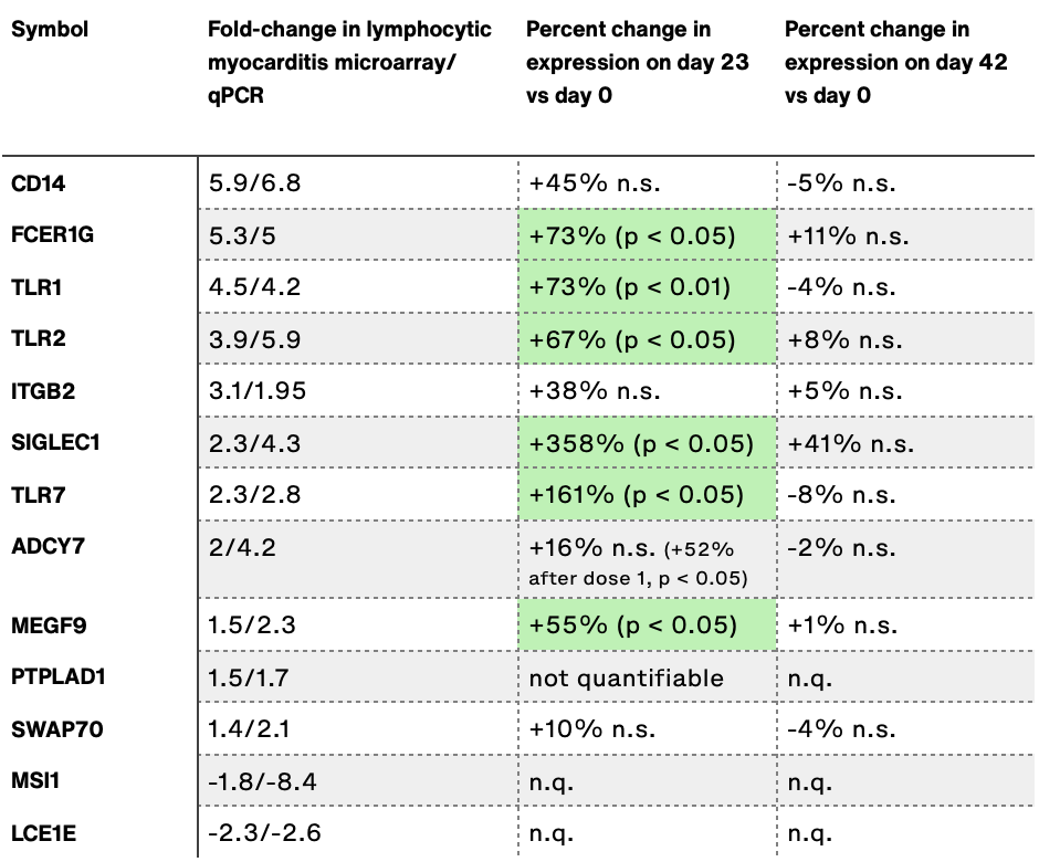
Note: Data in table is not normalized.
All 10 of the 10 quantifiable biomarkers were overexpressed, 6 of which significantly, indicating that myocarditis, possibly asymptomatic, may occur after vaccination. However, all of the upregulated genes appeared to normalize by day 42.
Discussion
Overall, the results show that vaccination with Comirnaty adversely affects almost every inspected biological pathway, but also that many effects subsided by day 42 post-vaccination. Most concerning are the genes whose expression did not fully normalize by D42, namely IFNG, IL6, PRKCA, PF4, PTGS1, PTGS2, FABP5, HIF1A and sialoadhesin (SIGLEC1). It is also noteworthy that most of the discussed genes were upregulated, and almost none were downregulated. Only PRKCA was consistently downregulated, while IL6 was only down on D1. The results are in agreement with the clinical presentation of VAEs, both in terms of symptoms and duration, and they provide a solid foundation for further research into VAEs caused by the COVID-19 vaccines. Unfortunately I was unable to find reliable plasma mRNA biomarkers for osteoporosis in literature, so a link between osteoporosis and COVID-19 vaccination remains to be shown.
The discovery that many STAT transcription factors, especially STAT3 and STAT5 were highly dysregulated following vaccination provides more circumstantial for the spike/STRA6 model of COVID-19 pathogenicity, and importantly, no evidence to reject the model could be found. A promising experiment to validate the spike/STRA6 model would be to determine whether STRA6-null mice (which are viable) can be infected by SARS-CoV-2 and whether they will exhibit COVID-19 symptoms, or whether STRA6 siRNA protects wild type mice from COVID-19.
In light of the fact that no deaths attributable to COVID-19 occurred in both the intervention and control groups of the Comirnaty phase 2/3 trial (Polack, Thomas et al. 2020), which took place over 3.5 months with 43,448 participants, the actual odds of dying from COVID-19 for healthy people (i.e. healthy enough to not have been excluded from the clinical trial) appear to be
over 3.5 months and therefore less than
in a single year. On the other hand, vaccination will modulate gene expression in 100% of recipients, with the potential of causing irreparable harm in some recipients. While these results don’t speak to the vaccine’s effectiveness, they definitely call the safety of Comirnaty into question.
Bibliography
Arunachalam, P. S., M. K. D. Scott, T. Hagan, C. Li, Y. Feng, F. Wimmers, L. Grigoryan, M. Trisal, V. V. Edara, L. Lai, S. E. Chang, A. Feng, S. Dhingra, M. Shah, A. S. Lee, S. Chinthrajah, S. B. Sindher, V. Mallajosyula, F. Gao, N. Sigal, S. Kowli, S. Gupta, K. Pellegrini, G. Tharp, S. Maysel-Auslender, S. Hamilton, H. Aoued, K. Hrusovsky, M. Roskey, S. E. Bosinger, H. T. Maecker, S. D. Boyd, M. M. Davis, P. J. Utz, M. S. Suthar, P. Khatri, K. C. Nadeau and B. Pulendran (2021). "Systems vaccinology of the BNT162b2 mRNA vaccine in humans." Nature 596(7872): 410-416.
Awosanya, O. D., C. E. Dalloul, R. J. Blosser, U. C. Dadwal, M. Carozza, K. Boschen, M. J. Klemsz, N. A. Johnston, A. Bruzzaniti, C. M. Robinson, E. F. Srour and M. A. Kacena (2022). "Osteoclast-mediated bone loss observed in a COVID-19 mouse model." Bone 154: 116227.
Berry, D. C., H. Jin, A. Majumdar and N. Noy (2011). "Signaling by vitamin A and retinol-binding protein regulates gene expression to inhibit insulin responses." Proc Natl Acad Sci U S A 108(11): 4340-4345.
Carpenter, R. L. and H.-W. Lo (2014). "STAT3 Target Genes Relevant to Human Cancers." Cancers 6(2): 897-925.
Dentelli, P., A. Trombetta, G. Togliatto, A. Zeoli, A. Rosso, B. Uberti, F. Orso, D. Taverna, L. Pegoraro and M. F. Brizzi (2009). "Formation of STAT5/PPARγ Transcriptional Complex Modulates Angiogenic Cell Bioavailability in Diabetes." Arteriosclerosis, Thrombosis, and Vascular Biology 29(1): 114-120.
Engblom, D., J.-W. Kornfeld, L. Schwake, F. Tronche, A. Reimann, H. Beug, L. Hennighausen, R. Moriggl and G. Schütz (2007). "Direct glucocorticoid receptor-Stat5 interaction in hepatocytes controls body size and maturation-related gene expression." Genes & development 21(10): 1157-1162.
Giannakodimos, I., G. V. Gkountana, D. Lykouras, K. Karkoulias and S. Tsakas (2021). "The Role of Interleukin-6 in the Pathogenesis, Prognosis and Treatment of Severe COVID-19." Curr Med Chem 28(26): 5328-5338.
Heidecker, B., M. M. Kittleson, E. K. Kasper, I. S. Wittstein, H. C. Champion, S. D. Russell, R. H. Hruban, E. R. Rodriguez, K. L. Baughman and J. M. Hare (2011). "Transcriptomic biomarkers for the accurate diagnosis of myocarditis." Circulation 123(11): 1174-1184.
Höpfinger, A., M. Berghoff, T. Karrasch, A. Schmid and A. Schäffler (2021). "Systematic Quantification of Neurotrophic Adipokines RBP4, PEDF, and Clusterin in Human Cerebrospinal Fluid and Serum." The Journal of Clinical Endocrinology & Metabolism 106(5): e2239-e2250.
Hough, S., L. V. Avioli, H. Muir, D. Gelderblom, G. Jenkins, H. Kurasi, E. Slatopolsky, M. A. Bergfeld and S. L. Teitelbaum (1988). "Effects of hypervitaminosis A on the bone and mineral metabolism of the rat." Endocrinology 122(6): 2933-2939.
Liu, J., C. Nie, L. Xue, Y. Yan, S. Liu, J. Sun, M. Fan, H. Qian, H. Ying, L. Wang and Y. Li (2021). "Growth hormone receptor disrupts glucose homeostasis via promoting and stabilizing retinol binding protein 4." Theranostics 11(17): 8283-8300.
Mahmoud, E., H. Tamer, A.-A. Yousry Esam-Eldin, S. Heba, M. S. Israa, F. A. E. M. Mohammed and A. Amr (2021). "STRA6, as A Novel Binding Receptor of COVID-19, A Breakthrough That could Explain COVID-19 Symptoms with Unknown Aetiology." Research Square.
Marwarha, G., D. C. Berry, C. M. Croniger and N. Noy (2014). "The retinol esterifying enzyme LRAT supports cell signaling by retinol-binding protein and its receptor STRA6." The FASEB Journal 28(1): 26-34.
Nan, J., Y. Wang, J. Yang and G. R. Stark (2018). "IRF9 and unphosphorylated STAT2 cooperate with NF-κB to drive IL6 expression." Proceedings of the National Academy of Sciences of the United States of America 115(15): 3906-3911.
Palma-Barqueros, V., N. Bohdan, N. Revilla, V. Vicente, J. M. Bastida and J. Rivera (2021). "PTGS1 gene variations associated with bleeding and platelet dysfunction." Platelets 32(5): 710-716.
Park, K. W., C. Y. Lin, E. N. Benveniste and Y. S. Lee (2016). "Mitochondrial STAT3 is negatively regulated by SOCS3 and upregulated after spinal cord injury." Exp Neurol 284(Pt A): 98-105.
Polack, F. P., S. J. Thomas, N. Kitchin, J. Absalon, A. Gurtman, S. Lockhart, J. L. Perez, G. Pérez Marc, E. D. Moreira, C. Zerbini, R. Bailey, K. A. Swanson, S. Roychoudhury, K. Koury, P. Li, W. V. Kalina, D. Cooper, R. W. Frenck, L. L. Hammitt, Ö. Türeci, H. Nell, A. Schaefer, S. Ünal, D. B. Tresnan, S. Mather, P. R. Dormitzer, U. Şahin, K. U. Jansen and W. C. Gruber (2020). "Safety and Efficacy of the BNT162b2 mRNA Covid-19 Vaccine." New England Journal of Medicine 383(27): 2603-2615.
Pretorius, E., M. Vlok, C. Venter, J. A. Bezuidenhout, G. J. Laubscher, J. Steenkamp and D. B. Kell (2021). "Persistent clotting protein pathology in Long COVID/Post-Acute Sequelae of COVID-19 (PASC) is accompanied by increased levels of antiplasmin." Cardiovascular Diabetology 20(1): 172.
Radominska-Pandya, A., G. Chen, P. J. Czernik, J. M. Little, V. M. Samokyszyn, C. A. Carter and G. Nowak (2000). "Direct interaction of all-trans-retinoic acid with protein kinase C (PKC). Implications for PKC signaling and cancer therapy." J Biol Chem 275(29): 22324-22330.
Schultz, N. H., I. H. Sørvoll, A. E. Michelsen, L. A. Munthe, F. Lund-Johansen, M. T. Ahlen, M. Wiedmann, A.-H. Aamodt, T. H. Skattør, G. E. Tjønnfjord and P. A. Holme (2021). "Thrombosis and Thrombocytopenia after ChAdOx1 nCoV-19 Vaccination." The New England journal of medicine 384(22): 2124-2130.
Shi, T., R. S. Papay and D. M. Perez (2012). "α(1A)-adrenergic receptor differentially regulates STAT3 phosphorylation through PKCϵ and PKCδ in myocytes." Journal of receptor and signal transduction research 32(2): 76-86.
Ullah, I., S. Paul, Z. Hong and Y. G. Wang (2019). "Significance tests for analyzing gene expression data with small sample sizes." Bioinformatics 35(20): 3996-4003.
Zhu, M., S. John, M. Berg and W. J. Leonard (1999). "Functional association of Nmi with Stat5 and Stat1 in IL-2- and IFNgamma-mediated signaling." Cell 96(1): 121-130.