
New Insights Into Vitamin A Metabolism


Key points:
- The vitamin A metabolite 4-oxo-retinoic acid (4-oxo-RA) is chemically and functionally similar to the naturally occurring 5-oxo-eicosapentaenoic acid (5-oxo-EPE)
- The C20:5 fatty acid EPE is an integral component of cell membranes that is released either mechanistically through physical damage to the membrane or enzymatically in response to hypoxia
- 5-oxo-EPE is a chemoattractant for immune cells and must be reduced to (5R)-OH-EPE before the immune response is stopped
- Retinoic acid (RA) is likely metabolized to mostly (4S)-hydroxy-RA [(4S)-OH-RA] by prostaglandin G/H synthase 1 (PTGS1), which is the target of nonsteroidal anti-inflammatory drugs (NSAIDs), in a chemical reaction highly similar to the formation of (5S)-OH-EPE by polyunsaturated fatty acid 5-lipoxygenase (ALOX5)
- Expression of PTGS1 was mildly correlated with expression of signal transducer and activator of transcription 3 (STAT3), strongly correlated with STAT5B and strongly inversely correlated with both STAT5A and peroxisome proliferator-activated receptor alpha (PPARA)
- It is likely that very-long-chain 3-oxoacyl-CoA reductase (HSD17B12) catalyzes the reduction of 4-oxo-RA to (4R)-OH-RA, which is then potentially metabolized to a substance with estrogen-like activity by cytochrome P450 2U1 (CYP2U1) and aromatase (CYP19A1), that is finally inactivated by corticosteroid 11-beta-dehydrogenase isozyme 2 (HSD11B2)
- Expression of HSD17B12 was highly correlated with expression of lipogenic genes, and inversely correlated with pro-inflammatory genes
- Retinol dehydrogenase 10 (RDH10) and the unexplored hydroxysteroid dehydrogenase-like protein 2 (HSDL2) are the best candidates for oxidizing retinol in vivo, while aldo-keto reductase family 1 member B10 (AKR1B10) and dehydrogenase/reductase SDR family member 9 (DHRS9) are the best candidates for reducing retinal
- Expression of STAT5A, which is activated by retinol, was correlated with many of the most downregulated genes in obesity, and additionally with PPARA, PPARG and epidermal growth factor receptor (EGFR), while expression of STAT5B was correlated with PTGS1, retinoic acid receptor alpha (RARA) and retinoic acid receptor RXR-beta (RXRB)
- In addition to receptor for retinol uptake STRA6 (STRA6)/STAT5, it appears that retinol also activates the glucocorticoid receptor (NR3C1)
Methods
I analyzed the expression of various genes in adipose tissue samples (Civelek, Wu et al. 2017). These samples are from a subset of METSIM study participants, which was a prospective population-based study of 10,197 Finnish men related to metabolic syndrome and cardiovascular diseases. Importantly, all the samples were from people believed to be healthy. I categorized the samples into the following groups:
- Control: all participants with BMI < 22.5 (n = 79)
- BMI 20–22.5 (n = 76); I don’t remember why I added this group, since it’s almost identical the control group (only 3 people had BMI < 20)
- BMI 22.5–25 (n = 180)
- BMI 25–27.5 (n = 240)
- BMI 27.5–30 (n = 161)
- BMI 30–32.5 (n = 67)
- BMI 32.5–35 (n = 30)
The groups seem large enough to be confident in the significance and precision of the results. The expression of genes in every group was compared to the control group. I chose to group the samples by BMI based on the assumption that vitamin A causes weight gain, due to the fact that vitamin A deficiency has been linked to weight loss in animals (Raja Gopal Reddy, Pavan Kumar et al. 2016), reviewed by (Chen 2021), but this assumption does not need to hold true for the results to be accurate. Correlations between genes were calculated irrespective of group membership for all test samples combined (i.e. BMI 22.5–35).
Gene expression was calculated using an app (ESGS) developed for this purpose. Results are expressed as percent change in expression of the gene in a test group compared to the expression of the same gene in the control group. For example, a value of 0% is equivalent to a fold-change of 1 or a log2(fold-change) of 0, and a value of -50% is equivalent to a fold-change of 0.5 or a log2(fold-change) of -1. Zero or negative values were excluded and assumed to be non-quantifiable. Error bars in the charts generated by ESGS denote the standard deviation. For comparison, the charts generated by ESGS also include microarray data from vitamin A-deficient (VAD) rat liver samples compared to vitamin A-sufficient (VAS) rat liver samples, n = 7 (McClintick, Crabb et al. 2006) and RNAseq from mouse small intestine samples, n = 3 (Chai, Lyu et al. 2020).
Significance of the results was determined for each group versus control, and additionally for each group versus all other test groups, using Welch’s t test, which was reported to be reliable for this purpose (Ullah, Paul et al. 2019). (*), (**) and (***) denote a significant change versus control with p < 0.05, p < 0.01 and p < 0.001, respectively whereas (†), (††) and (†††) denote a significant change versus the remaining test groups with p < 0.05, p < 0.01 and p < 0.001 respectively.
Curve fitting, including calculation of R2 of the results was performed using Apple’s Numbers app using the calculated results (percent change). Correlations were established using ESGS by calculating the Pearson correlation coefficient for raw sample-level data, including zero values for samples in which no mRNA could be detected.
Lipogenic genes are progressively downregulated with BMI
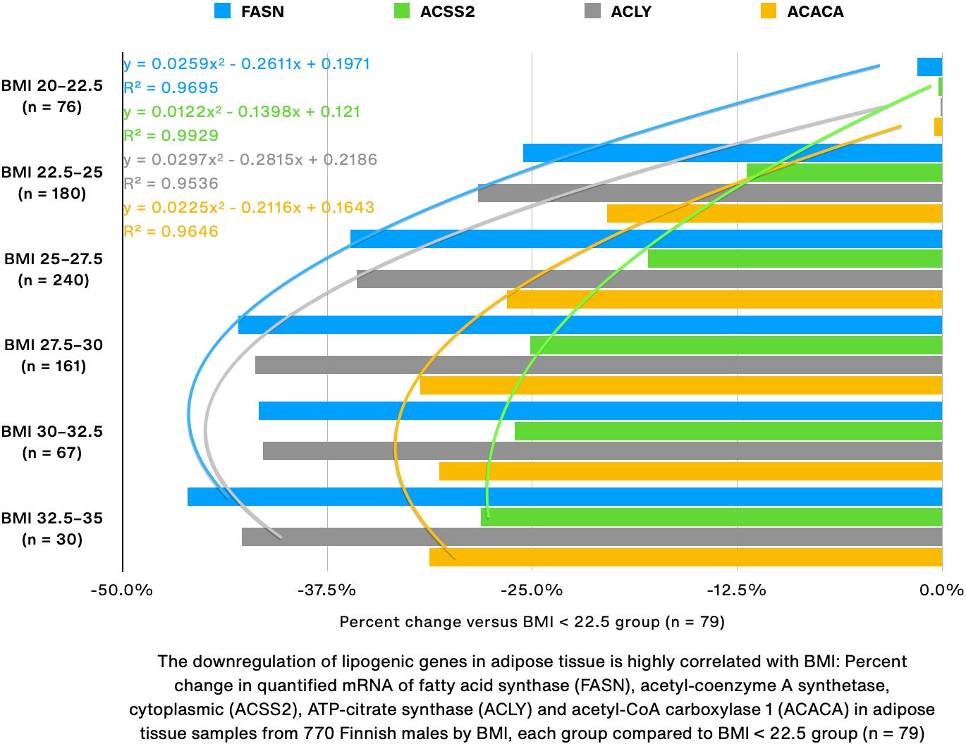
Since I had been reading a study about fatty acid metabolism in cancer (Kuo and Ann 2018) I decided to first look at the expression of lipogenic genes. Surprisingly, all four of the tested lipogenic genes were progressively more downregulated as BMI increased (-28% to -46% at BMI 32.5–35, p < 0.001), which is not what would be expected in overweight and obese subjects. Even more surprising are the R2 values calculated by Numbers (0.95–0.993), which are a measure of how good the curves fit to the data (1 is the best). According to Google, in finance an R2 value of 0.7 is considered good, and in academic research, an R2 of 0.75 is considered “substantial”.
Expression of STAT3, STAT5A and PPARA was correlated
Next, I tried to determine how weight gain is even possible if all the lipogenic genes are downregulated, by comparing the expression of peroxisome proliferator-activated receptor alpha (PPARA), which induces fatty acid oxidation, signal transducer and activator of transcription 5A (STAT5A), which is activated directly by Vitamin A and induces lipogenesis, and STAT3, which repairs damage to membranes and cells and is not directly activated by Vitamin A but likely activated by damage caused by Vitamin A. I did not include PPAR gamma (PPARG), which induces lipogenesis, because it was found to be highly correlated with PPARA (data not shown).
Again, there is an almost perfect correlation between all three genes. Notably STAT3 consistently increases, indicating that there is progressively more damage that needs to be repaired as BMI increases. Additionally STAT5A was downregulated just a tiny bit more than PPARA in almost every range.
Expression of PPARA, RDH10, HSD17B12, CYP2U1 and HSD11B2 was correlated
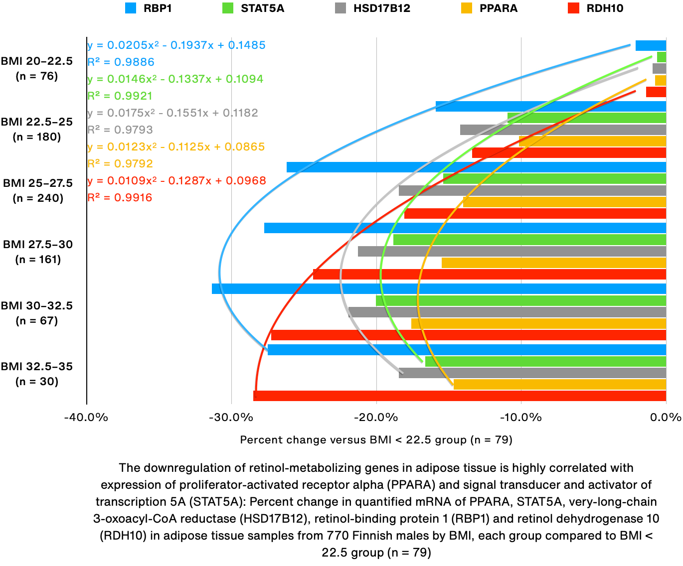
I then began looking for other genes with the same distribution pattern as PPARA/STAT5A. A number of genes were correlated with PPARA and STAT5A, namely very-long-chain 3-oxoacyl-CoA reductase (HSD17B12), retinol-binding protein 1 (RBP1), retinol dehydrogenase 10 (RDH10), cytochrome P450 2U1 (CYP2U1) and corticosteroid 11-beta-dehydrogenase isozyme 2 (HSD11B2; data not shown for CYP2U1 and HSD11B2).
Interestingly, of all 17β-hydroxysteroid dehydrogenases (HSD17Bs) none correlated with STAT5A as well as HSD17B12, which has not been reported to be active towards retinol. However, since the correlation is so strong, and because of its known activity towards hormones, I believe that HSD17B12 could be one of the primary enzymes responsible for metabolizing retinol in humans. It catalyzes the reduction of estrone’s oxo group to form 17β-estradiol, which I believe is really (17R)β-estradiol, and additionally, it was recently shown to reduce the carcinogenic 4-(methylnitrosamino)-1-(3-pyridyl)-1-butanone (NNK) from tobacco smoke to 4-(methylnitrosamino)-1-(3-pyridyl)-1-butanol (NNAL) in human lungs (Ashmore, Luo et al. 2018). Notably, 95% of the product formed was (R)-NNAL, and just 5% was (S)-NNAL. This enzyme is therefore one of the best candidates for reducing either 4-oxo-RA to (4R)-OH-RA or 4-oxo-retinol to (4R)-OH-retinol.
RDH10 oxidizes free retinol (i.e. not bound to RBP1) to retinal, with a strong preference for all-trans-retinol over 9-cis-retinol and 11-cis-retinol. Its downregulation is possibly not related to substrate availability and could be a result of either PPAR, STAT or other transcription factor activity.
HSD11B2 catalyzes the inactivation of cortisol (11β-hydrocortisone) by oxidation to cortisone (11β-oxo-cortisone), and it is inhibited by licorice. While its role in retinol metabolism remains unclear, it is possible that HSD11B2 oxidizes hydroxylated retinol or RA to 4-oxo-retinol and 4-oxo-RA, respectively, contributing to the feedback loop in which 4-oxo-RA is not eliminated. On the other hand, its affinity for the 11th carbon in steroids suggests that it is unlikely to be active towards the fourth carbon in retinoids.
Finally, CYP2U1 is by far the most interesting of the tested enzymes due to its involvement in inflammation and the arachidonic acid signaling pathway. Recall that of all the noncyclical fatty acids, retinoic acid is most chemically similar to arachidonic acid (AA; specifically eicosapentaenoic acid, EPE), since both RA and EPE are carboxylic acids, have 20 carbon atoms and five unsaturated double bonds. I believe the term EPE specifically refers to the all-cis configuration (5Z,8Z,11Z,14Z,17Z), whereas RA is either arranged with one (all-trans-RA) or two (9-cis-RA and 11-cis-RA) cis-double bonds, the first of which always being at the fifth carbon of the cyclohexene ring. The recently identified endogenous retinoic acid receptor RXR (RXR) ligand 9-cis-13,14-dihydroretinoic acid (9CDHRA) is more similar to eicosatetraenoic acid (ETE), as both 9CDHRA and ETE have four unsaturated double bonds. The AA signaling pathway is activated in response to hypoxia, cellular insult or general stress and causes inflammation until the damage is resolved and it is inactivated. In my opinion, understanding the AA signaling pathway is crucial to understanding the effects of Vitamin A on the body.
The arachidonic acid signaling pathway
In general, when a cell is under stress and activates the AA pathway, the following events happen (adapted from Wikipedia):
- Phospholipase A2 (PLA2) enzymes are activated and release all-trans-eicosapentaenoic acid (atEPE) from membrane phospholipids; atEPE is an integral component of cell membranes, and removing it not only weakens the membrane but signals to the immune system that the cell is in trouble and needs help
- Polyunsaturated fatty acid 5-lipoxygenase (ALOX5) catalyzes oxygenation of atEPE to 5(S)-hydroperoxy-EPE, which is a radical very alike to 4-hydroperoxy-RA
- Cellular peroxidases rapidly reduce 5(S)-hydroperoxy-EPE to 5(S)-OH-EPE
- 5(S)-OH-EPE is oxidized to 5-oxo-EPE, which is 30–100x more potent (according to Wikipedia), by a 5-hydroxyeicosanoid dehydrogenase (5-HEDH), whose activity has been demonstrated in humans in vivo but for which no gene has yet been identified
Importantly, the fourth step in the pathway does not happen under normal physiological conditions, because it requires NADP+ as a co-factor, which is not present in large enough quantities in cells under normoxia. It is however present in sufficiently large quantities after cellular insult and the resulting hypoxia. 5-oxo-ETE (which has one less double bond than 5-oxo-EPE) directly induces an immune response by activating oxoeicosanoid receptor 1 (OXER1):
“5-Oxo-ETE is a potent chemoattractant for eosinophils and has similar effects on neutrophils, basophils and monocytes. It elicits infiltration of eosinophils and, to a lesser extent, neutrophils into the skin after intradermal injection in humans. It also promotes the survival of tumor cells and has been shown to block the induction of apoptosis by 5-LO inhibitors.” (Grant, Rokach et al. 2009)
However, a different study found that 5-oxo-ETE did not promote the survival of tumor cells and instead arrested growth and induced apoptosis in all four of the tested cancer cell lines (O'Flaherty, Rogers et al. 2005). 5-oxo-15-OH-ETE, but importantly not 5-OH-ETE, produced the same results. It should be investigated whether 4-oxo-RA is an OXER1 receptor ligand.
In any case, the sequence of events ends either when the insulted cell is destroyed by immune cells, or when NADPH is restored, which prevents the formation of 5-oxo-EPE. The AA pathway is usually activated by the body directly in response to stress, such as bacterial infection, however it can also be induced by xenobiotics, with one crucial difference in Step 2:
- Cytochrome P450 enzymes, and not ALOX5, catalyze the formation of (5R)-OH-EPE instead of (5S)-OH-EPE
CYP2U1 normally terminates inflammatory signaling through the AA pathway by hydroxylating the tail of EPE at either the 20th or 19th carbon, which in the nomenclature of retinoic acid would translate to the 16th or 17th carbon. CYP2U1 was reported to metabolize many fatty acids including ETE (C20:4) and EPE (C20:5) (Chuang, Helvig et al. 2004), but its activity towards 5-hydroxylated fatty acids is unclear. Its expression was highly correlated with the other retinol-related genes, which suggests that it plays some kind of role in metabolizing retinol, possibly oxidizing 4-OH-RA to 4,16-(OH)2-RA, 4-oxo-RA to 4-oxo-16-OH-RA, RA to 16-OH-RA or retinol to 16-OH-retinol, however it is unlikely to be involved in the biosynthesis of 4- or 18-hydroxylated retinol derivatives like 4-oxo-RA and 4-oxo-18-OH-RA. If RA behaves similar to EPE, that would suggest that hydroxylation at the 16 position is required to inactivate RA and that hydroxylation at the 18 position promotes the inflammatory cycle.
PTGS1 could be the most important enzyme in the biotransformation of retinol
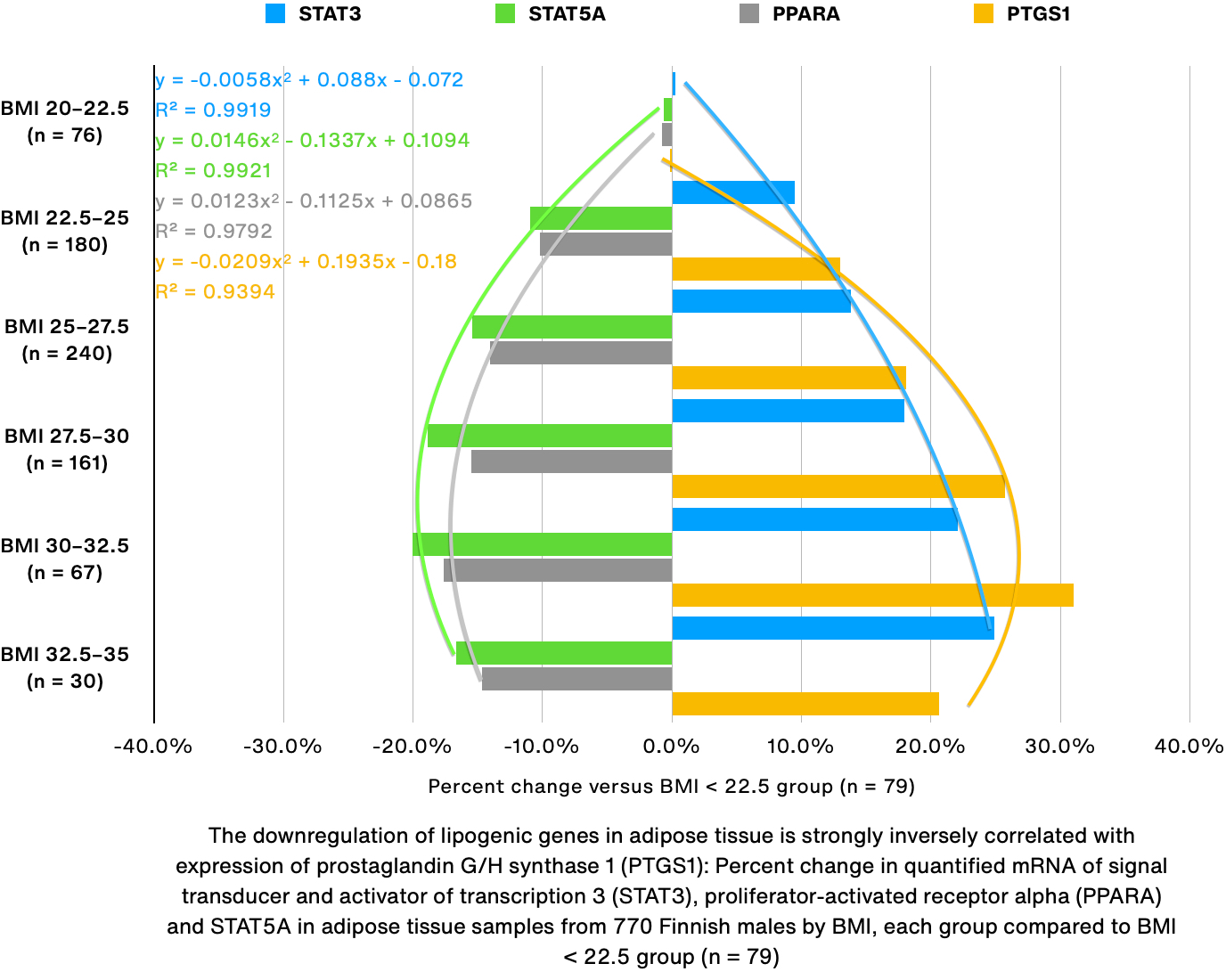
While looking at various CYP enzymes to determine which is primarily responsible for forming 4-OH-RA, I remembered the involvement of prostaglandin G/H synthase 1 (PTGS1). Specifically, “RA undergoes hydroperoxide (H2O2 or PPHP)- or arachidonic acid-dependent, PGH synthase-catalyzed metabolism as evidenced by ultraviolent [sic] spectroscopic analysis of reaction mixtures” (Samokyszyn, Chen et al. 1995). Furthermore, the formation of 4-OH-RA described by Samokyszyn et. al. is virtually identical to the biotransformation of EPE in the AA singling pathway, since it involves formation of either a 4-hydroperoxy or a 5,6-epoxy radical. Most importantly, I discovered that expression of PTGS1 was weakly correlated with STAT3 and strongly inversely correlated with PPARA and STAT5A (see figure on previous page).
Strangely, the sum of all quadratic coefficients just barely exceeded zero:
I haven't figured out the significance of this, but I feel like the number 0.0002 is too close to zero for this to just be a random statistical effect.
In any case, the ratio of 4-OH-RA enantiomers produced by PTGS1 has not ben experimentally determined, but it was shown to prefer forming (15S)-OH-ETE over (15R)-OH-ETE with a 15S/15R ratio of 7:3, or 2.3x greater production of (15S)-OH-ETE (Johnsson, Rönnberg et al. 2021), making it likely that PTGS1 preferentially forms (4S)-OH-RA. The reason for this could very plausibly be that PTGS1 has no known exogenous ligands (except atRA), and transformation of atRA by PTGS1 would therefore signal to other cells that damage to cell membranes has occurred, even though damage did not actually occur. It is also plausible that such a situation would elicit an immune response even if there was no actual damage, and that the only real damage is subsequently caused by the immune response and modulation of gene expression.
PTGS1-null mice do not exhibit any major abnormalities except reduced inflammatory response after topical challenge with ETE, and decreased platelet activation, which I believe leads to increased bleeding and decreased clotting (Palma-Barqueros, Bohdan et al. 2021). Therefore, even though RA has been reported to damage cell membranes in vitro (Shih 1997), it now appears possible that retinoic acid doesn’t directly cause this in vivo (because PTGS1-null mice showed no abnormalities), probably because RA immediately associates with transport proteins like albumin (ALB), cellular retinoic acid-binding proteins (RABPs) and fatty acid-binding proteins (FABPs). It also appears that toxicity is instead caused by immune cells recruited after progressive oxidation of free retinol to (4S)-OH-RA by PTGS1. The immune response is likely attenuated by formation of the (4R)-OH-RA enantiomer.
Furthermore, all available nonsteroidal anti-inflammatory drugs (NSAIDs) inhibit PTGS1 and PTGS2 with different affinities, notably flurbiprofen and ketoprofen specifically inhibit PTGS1, ibuprofen (Advil) and naproxen (Aleve) inhibit both PTGS1 and PTGS2, and diclofenac inhibits PTGS2 (Cryer and Feldman 1998). It is possible that NSAIDs reduce inflammation by preventing pro-inflammatory metabolism of retinoic acid by PTGS. It is also possible that dietary retinol increases the risk of blood clots by increasing expression of PTGS1, although my results are only from adipocytes.
Additional verification of the correlations
In order to mathematically verify the visually apparent correlations, I updated ESGS to calculate the Pearson correlation coefficient (r) for the tested genes. I believe that this value is only meaningful for genes with 100% quantifiability (because otherwise the zero values interfere with the correlation), which notably excludes RBP1, receptor for retinol uptake STRA6 (STRA6), aldo-keto reductase family 1 member B10 (AKR1B10) and more. I also updated ESGS to calculate the Top 25 most up- and downregulated genes (of all 18,000 genes tested, excluding those with <75% quantifiability), in addition to the Top 25 most correlated and inversely correlated genes for any query gene.
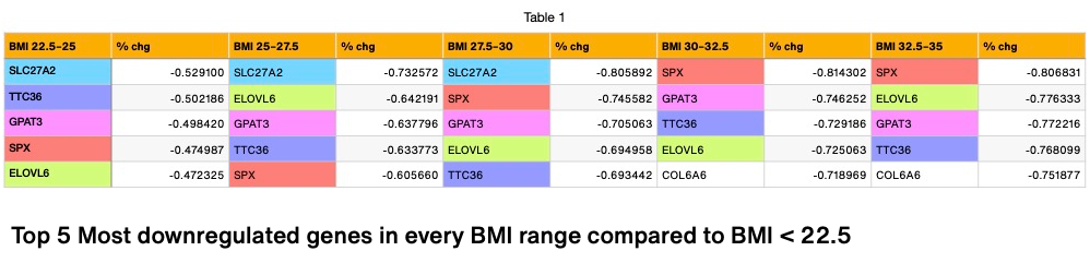
Interestingly, STAT5A was correlated with 3 of the 5 most downregulated genes overall, namely tetratricopeptide repeat protein 36 (TTC36; r = 0.355), glycerol-3-phosphate acyltransferase 3 (GPAT3; 0.378) and spexin (SPX; 0.432), in addition to PPARA (0.665), PPARG (0.537), retinoic acid receptor RXR-beta (RXRB; 0.247), HSD17B12 (0.285), RDH10 (0.407) and epidermal growth factor receptor (EGFR; 0.477). It was also inversely correlated with PTGS1 (-0.223) and platelet-activating factor acetylhydrolase (PLA2G7; -0.337).The correlation between STAT5B and these genes was usually weaker or absent, except for RXRB (0.287), retinoic acid receptor alpha (RARA; 0.567), EGFR (-0.288) and PTGS1 (0.464). It is unclear why the r value for EGFR is negative for STAT5A and positive for STAT5B. STAT3 was found to be inversely correlated with PPARG (-0.409), CYP2U1 (-0.402) and RDH10 (-0.507). Furthermore, CYP2U1 was found to correlate with RARA (-0.53), PPARG (0.37) and HSD17B12 (0.32).
I then tried to identify transcription factors for retinol-related genes. The transcription factor with the highest correlation to RDH10 was determined to be the glucocorticoid receptor (NR3C1; r = 0.62), whose endogenous ligands are cortisol, cortisone and aldosterone, that is pharmacologically activated by the steroid dexamethasone and inhibited by ketoconazole. Additionally, it has been reported to function as a co-activator for transcription of STAT5 target genes. Of all genes, the #3 most correlated gene with RDH10 was hydroxysteroid dehydrogenase-like protein 2 (HSDL2; r = 0.649), which has not been investigated much but appears theoretically capable of oxidizing retinol.
Interestingly, of all genes, HSD17B12 was most correlated with acetyl-CoA carboxylase 1 (ACACA; r = 0.669), which catalyzes the rate-limiting step in fatty acid synthesis, and it was also highly correlated with NADH-ubiquinone oxidoreductase 75 kDa subunit, mitochondrial (NDUFS1; r = 0.605), a mitochondrial enzyme that catalyzes the oxidation of NADH during respiration. In fact, none of the Top 25 genes most positively correlated with HSD17B12 were transcription factors, and most of them were related to mitochondrial beta-oxidation. Unexpectedly, the gene most inversely correlated with HSD17B12 was herpesvirus entry mediator (TNFRSF14; r = -0.425), and many of the other inversely correlated genes were related to immune response and membrane lipids, for example retinoic acid receptor responder protein 3 (PLAAT4; -0.405), mitogen-activated protein kinase kinase kinase 3 (MAP3K3; -0.394) and phosphoinositide 3-kinase regulatory subunit 5 (PIK3R5; -0.391).
The transcription factor most correlated with CYP2U1 was GDNF-inducible zinc finger protein 1 (GZF1; 0.678), which has been linked to morphogenesis and Larsen syndrome. Interestingly, of all genes, the circular RNA septin-9 (SEPTIN9) was most inversely correlated with CYP2U1 (-0.701), and SEPTIN9 was reported to be a target of transcription factor E2F1 (Zheng, Huang et al. 2020).
Finally, PTGS1 was extremely correlated with a number genes, all of which I’m not too familiar with, most prominently acrosin-binding protein (ACRBP; r = 0.911), platelet factor 4 (PF4; 0.877), tetraspanin-33 (TSPAN33; 0.874) and small membrane A-kinase anchor protein (C2orf88; 0.873). Additionally, PTGS1 was inversely correlated with alpha-aminoadipic semialdehyde dehydrogenase (ALDH7A1; -0.488). PF4-antibodies have been linked to blood clots after vaccination with Vaxzevria, COVID-19 Vaccine (ChAdOx1-S [recombinant]) (Schultz, Sørvoll et al. 2021).
Proposed biotransformation of retinol
Based on the data, I propose the following biotransformation of retinol:
- Retinol is oxidized to retinal by RDH10, HSDL2 and others
- Retinal is either oxidized to retinoic acid by retinal dehydrogenases (ALDH1As), or reduced back to retinol by dehydrogenase/reductase SDR family member 9 (DHRS9), AKR1B10 and others
- Retinoic acid is oxidized—producing free radical intermediates (Samokyszyn, Chen et al. 1995)—in highly variable ratios, to (4S)-OH-RA and (4R)-OH-RA, by specific and non-specific enzymes, the most important enzymes likely being PTGS1 (7:3 4S/4R ratio); of the CYP enzymes CYP1A1, CYP26B1 and CYP3A4 (1:1 ratio each) and CYP26C1 and CYP2C9 (both preferring 4R) (Shimshoni, Roberts et al. 2012)
- Under normoxia, one or possibly both 4-OH-RA enantiomers can be conjugated by UGT2B7 (Samokyszyn, Gall et al. 2000) and possibly UGT1A10 (Trivedi, Gilliland et al. 2006), resulting in their elimination, or further oxidized to 4,16-(OH)2-RA by CYP2U1, CYP26s and others, however it is unclear how much 4-oxo-RA is formed under normoxia
- 4-OH-RA glucuronide is eliminated in feces
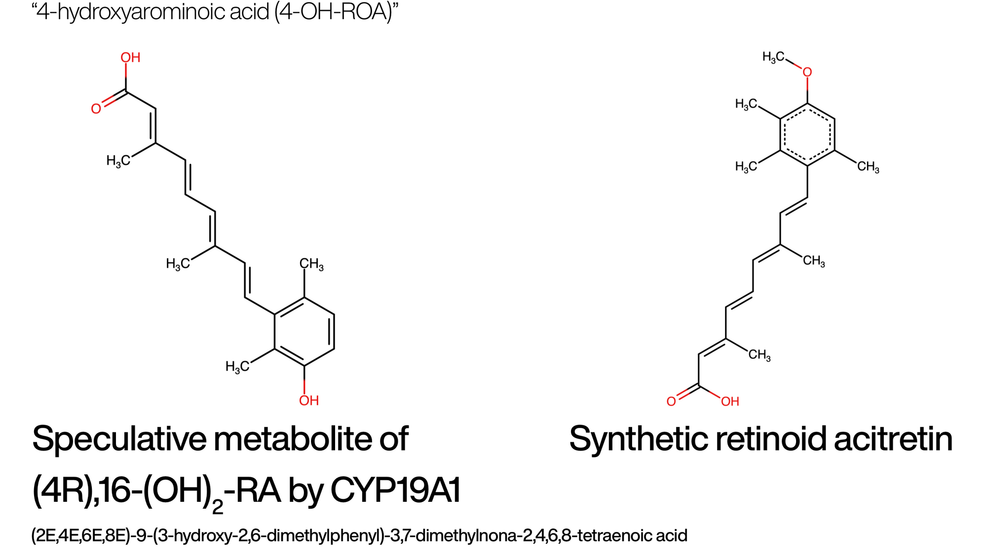
- 4,16-(OH)2-RA may undergo aromatization by CYP19A1 to a substance similar to the synthetic retinoid acitretin, which I have named “arominoic acid” (ROA), with “arominol” (ROOL; portmanteau of “aromatic” and “retinol”) being the corresponding alcohol; the product formed by CYP19A1 would therefore be called (4R)-OH-ROA; expression of CYP19A1 was not correlated with STAT5A as much as the other genes but increases monotonically with BMI and is up at BMI 32.5–35 (+118%; p < 0.001), down in VAD rat liver samples (-50%; p < 0.05; n = 7) and off in VAD mouse small intestine samples (n = 3); it seems like 4-OH-ROA would be active as an estrogen, since aromatization of testosterone (which contains a cyclohexene ring) by CYP19A1 produces estradiol (which contains a benzene ring); is is possible that in a subsequent reaction HSD11B2 inactivates ROA by oxidizing the hydroxy group at the four position to form 4-oxo-ROA
- Under hypoxia, substantial amounts of retinoic acid are oxidized to (4S)-OH-RA and subsequently to 4-oxo-RA, and 4-oxo-RA is reduced to (4R)-OH-RA by HSD17B12; this cycle continues indefinitely until normoxia is restored or the cell undergoes apoptosis
Discussion
It is now no longer difficult to imagine how retinoic acid could cause auto-immune disease, since the data appears to confirm that retinol is effectively a pro-inflammatory proto-cytokine that is stored in the liver and, once the liver is saturated, in different types of tissue all over the body, especially in epithelial cells that constitute blood–tissue barriers, and in adipocytes, both of which were shown to express STRA6 (Amengual, Zhang et al. 2014).
To give an example, let me point out the pathogenesis of acne. It is believed that acne is caused by the opportunistic bacterium C. acnes, which resides in hypoxic areas of the skin and metabolizes cobalamin (vitamin B12). It has previously been suggested that C. acnes normally biosynthesizes vitamin B12, but when vitamin B12 from the host becomes available to it, it transforms it into porphyrins, which have been implicated by investigators as a causal factor for the inflammation in acne (Kang, Shi et al. 2015). However, it has also been shown that C. acnes synthesizes lipolytic enzymes, and that it derives energy from lipase-mediated sebum lipid degradation (Kim, Lee et al. 2020).
I propose that C. acnes is not pathogenic at all, and that it mainly causes inflammation not by synthesizing porphyrins, but instead by hydrolyzing retinyl esters, which are abundant in epithelial cells and possibly in sebum itself. The initial liberation of retinol and subsequent oxidation to retinoic acid attracts immune cells to the region, which proceed to attack C. acnes. During this attack, which causes oxidative stress and damages healthy cells in the vicinity, even more retinol is liberated from surrounding cells, resulting in a detrimental cycle that is difficult to stop. In fact, it seems to me that one strategy to potentially halt this chain of events is the formation of scar tissue (fibrogenesis), which I would imagine is impenetrable to lipids and retinoic acid. This would explain not only scarring in skin diseases but also for example scarring of the liver in non-alcoholic steatohepatitis (NASH).
It is also no longer difficult to imagine how retinoic acid could directly cause carcinogenesis. From an evolutionary standpoint, organisms and individual cells evolve when they are confronted with danger and must adapt to survive. Recall that the AA signaling pathway consists of a challenge (e.g. damage to membranes), a response which includes formation of an S-enantiomer, and a resolution which includes the formation of an R-enantiomer. When cells are challenged with retinoic acid, however, as described previously there is no simple resolution, and progressively more S-enantiomers are liberated. Since the pro-inflammatory cycle does not simply progress, but constantly escalates (as long as retinyl esters in the vicinity are hydrolyzed), progressively more evolutionary pressure is exerted on cells to resolve the challenge through mutation. It is therefore not difficult at all to imagine that, under that kind of pressure, and additionally in the presence of the ultimate morphogen retinoic acid, cells undergo transformation, which can also result in malignant transformations. Some circumstantial evidence for this theory is the dysregulation of many retinol-related genes in many different types of cancer, for example in non-small-cell lung cancer (Kuznetsova, Zinovieva et al. 2016). Additionally, atRA has been shown to induce micronucleation in human lymphocytes via chromosome breakage and other mechanisms (Alakhras, Stephanou et al. 2011).
The fact that introduction of additional exogenous retinoic acid is detrimental to cancer cells appears to add support to this theory. Since the transformed cells have only mutated to adapt to endogenous concentrations of retinoic acid, they are unable to defeat the secondary challenge with even more retinoic acid. However, if additional retinoic acid is administered for a long enough period time, exactly as is the case during some types of chemotherapy, it is only logical to expect that the transformed cells will undergo additional mutations to resist the secondary and subsequent challenges.
Bibliography
Alakhras, R. S., G. Stephanou, N. A. Demopoulos and S. S. Nikolaropoulos (2011). "Genotoxicity of all-trans retinoic acid (ATRA) and its steroidal analogue EA-4 in human lymphocytes and mouse cells in vitro." Cancer Lett 306(1): 15-26.
Amengual, J., N. Zhang, M. Kemerer, T. Maeda, K. Palczewski and J. Von Lintig (2014). "STRA6 is critical for cellular vitamin A uptake and homeostasis." Human molecular genetics 23(20): 5402-5417.
Ashmore, J. H., S. Luo, C. J. W. Watson and P. Lazarus (2018). "Carbonyl reduction of NNK by recombinant human lung enzymes: identification of HSD17β12 as the reductase important in (R)-NNAL formation in human lung." Carcinogenesis 39(8): 1079-1088.
Chai, Z., Y. Lyu, Q. Chen, C.-H. Wei, L. M. Snyder, V. Weaver, A. Sebastian, I. Albert, Q. Li, M. T. Cantorna and A. C. Ross (2020). "RNAseq studies reveal distinct transcriptional response to vitamin A deficiency in small intestine versus colon, uncovering novel vitamin A-regulated genes." bioRxiv: 798504.
Chen, G. (2021). "The Interactions of Insulin and Vitamin A Signaling Systems for the Regulation of Hepatic Glucose and Lipid Metabolism." Cells 10(8): 2160.
Chuang, S. S., C. Helvig, M. Taimi, H. A. Ramshaw, A. H. Collop, M. a. Amad, J. A. White, M. Petkovich, G. Jones and B. Korczak (2004). "CYP2U1, a Novel Human Thymus- and Brain-specific Cytochrome P450, Catalyzes ω- and (ω-1)-Hydroxylation of Fatty Acids *." Journal of Biological Chemistry 279(8): 6305-6314.
Civelek, M., Y. Wu, C. Pan, C. K. Raulerson, A. Ko, A. He, C. Tilford, N. K. Saleem, A. Stančáková, L. J. Scott, C. Fuchsberger, H. M. Stringham, A. U. Jackson, N. Narisu, P. S. Chines, K. S. Small, J. Kuusisto, B. W. Parks, P. Pajukanta, T. Kirchgessner, F. S. Collins, P. S. Gargalovic, M. Boehnke, M. Laakso, K. L. Mohlke and A. J. Lusis (2017). "Genetic Regulation of Adipose Gene Expression and Cardio-Metabolic Traits." Am J Hum Genet 100(3): 428-443.
Cryer, B. and M. Feldman (1998). "Cyclooxygenase-1 and cyclooxygenase-2 selectivity of widely used nonsteroidal anti-inflammatory drugs." Am J Med 104(5): 413-421.
Grant, G. E., J. Rokach and W. S. Powell (2009). "5-Oxo-ETE and the OXE receptor." Prostaglandins & other lipid mediators 89(3-4): 98-104.
Johnsson, A.-K., E. Rönnberg, D. Fuchs, J. Kolmert, J. Säfholm, H.-E. Claesson, M. Hamberg, C. E. Wheelock, G. Nilsson and S.-E. Dahlén (2021). "COX-1 dependent biosynthesis of 15-hydroxyeicosatetraenoic acid in human mast cells." Biochimica et Biophysica Acta (BBA) - Molecular and Cell Biology of Lipids 1866(5): 158886.
Kang, D., B. Shi, M. C. Erfe, N. Craft and H. Li (2015). "Vitamin B12 modulates the transcriptome of the skin microbiota in acne pathogenesis." Science translational medicine 7(293): 293ra103-293ra103.
Kim, H. J., B.-J. Lee and A.-R. Kwon (2020). "The grease trap: uncovering the mechanism of the hydrophobic lid in Cutibacterium acnes lipase[S]." Journal of Lipid Research 61(5): 722-733.
Kuo, C.-Y. and D. K. Ann (2018). "When fats commit crimes: fatty acid metabolism, cancer stemness and therapeutic resistance." Cancer communications (London, England) 38(1): 47-47.
Kuznetsova, E. S., O. L. Zinovieva, N. Y. Oparina, M. M. Prokofjeva, P. V. Spirin, I. A. Favorskaya, I. B. Zborovskaya, N. A. Lisitsyn, V. S. Prassolov and T. D. Mashkova (2016). "Abnormal expression of genes that regulate retinoid metabolism and signaling in non-small-cell lung cancer." Molecular Biology 50(2): 220-229.
McClintick, J. N., D. W. Crabb, H. Tian, J. Pinaire, J. R. Smith, R. E. Jerome and H. J. Edenberg (2006). "Global effects of vitamin A deficiency on gene expression in rat liver: evidence for hypoandrogenism." The Journal of Nutritional Biochemistry 17(5): 345-355.
O'Flaherty, J. T., L. C. Rogers, C. M. Paumi, R. R. Hantgan, L. R. Thomas, C. E. Clay, K. High, Y. Q. Chen, M. C. Willingham, P. K. Smitherman, T. E. Kute, A. Rao, S. D. Cramer and C. S. Morrow (2005). "5-Oxo-ETE analogs and the proliferation of cancer cells." Biochim Biophys Acta 1736(3): 228-236.
Palma-Barqueros, V., N. Bohdan, N. Revilla, V. Vicente, J. M. Bastida and J. Rivera (2021). "PTGS1 gene variations associated with bleeding and platelet dysfunction." Platelets 32(5): 710-716.
Raja Gopal Reddy, M., C. Pavan Kumar, M. Mahesh, M. Sravan Kumar, S. Mullapudi Venkata, U. K. Putcha, A. Vajreswari and S. M. Jeyakumar (2016). "Vitamin A deficiency suppresses high fructose-induced triglyceride synthesis and elevates resolvin D1 levels." Biochimica et Biophysica Acta (BBA) - Molecular and Cell Biology of Lipids 1861(3): 156-165.
Samokyszyn, V. M., T. Chen, K. R. Maddipati, T. J. Franz, P. A. Lehman and R. V. Lloyd (1995). "Free radical oxidation of (E)-retinoic acid by prostaglandin H synthase." Chem Res Toxicol 8(5): 807-815.
Samokyszyn, V. M., W. E. Gall, G. Zawada, M. A. Freyaldenhoven, G. Chen, P. I. Mackenzie, T. R. Tephly and A. Radominska-Pandya (2000). "4-Hydroxyretinoic Acid, a Novel Substrate for Human Liver Microsomal UDP-glucuronosyltransferase(s) and Recombinant UGT2B7 *." Journal of Biological Chemistry 275(10): 6908-6914.
Schultz, N. H., I. H. Sørvoll, A. E. Michelsen, L. A. Munthe, F. Lund-Johansen, M. T. Ahlen, M. Wiedmann, A.-H. Aamodt, T. H. Skattør, G. E. Tjønnfjord and P. A. Holme (2021). "Thrombosis and Thrombocytopenia after ChAdOx1 nCoV-19 Vaccination." The New England journal of medicine 384(22): 2124-2130.
Shih, T., Lin, T., Shealy, Y., & Hill, D. (1997). "Nonenzymatic isomerization of 9-CIS-Retinoic Acid catalyzed by Sulfhydryl Compounds." Drug Metabolism and Disposition 25(1): 27-32.
Shimshoni, J. A., A. G. Roberts, M. Scian, A. R. Topletz, S. A. Blankert, J. R. Halpert, W. L. Nelson and N. Isoherranen (2012). "Stereoselective formation and metabolism of 4-hydroxy-retinoic Acid enantiomers by cytochrome p450 enzymes." The Journal of biological chemistry 287(50): 42223-42232.
Trivedi, N. R., K. L. Gilliland, W. Zhao, W. Liu and D. M. Thiboutot (2006). "Gene Array Expression Profiling in Acne Lesions Reveals Marked Upregulation of Genes Involved in Inflammation and Matrix Remodeling." Journal of Investigative Dermatology 126(5): 1071-1079.
Ullah, I., S. Paul, Z. Hong and Y. G. Wang (2019). "Significance tests for analyzing gene expression data with small sample sizes." Bioinformatics 35(20): 3996-4003.
Zheng, X., M. Huang, L. Xing, R. Yang, X. Wang, R. Jiang, L. Zhang and J. Chen (2020). "The circRNA circSEPT9 mediated by E2F1 and EIF4A3 facilitates the carcinogenesis and development of triple-negative breast cancer." Mol Cancer 19(1): 73.